David Bovard - Science is beautiful
1. Why did you become a scientist?
I have always described myself as being very curious about everything that surrounds me. Even as a kid, I was always asking question and trying to understand how things were working. On top of that, I always said, as a kid, that I would one day become an "inventor" because I was always enthusiastic about finding new solutions or new ideas. One of the only jobs that offers you the opportunity to satisfy your scientific curiosity and at the same time to express your creativity is of course a scientist.

2. What aspects of science are beautiful to you?
The most striking aspect of science and therefore of the nature that surrounds us is certainly the incredible work done by natural selection that has pushed every element on earth to be perfectly optimized. Every day, I find myself amazed by the mechanisms put in place by nature that allow simple or living organisms to survive at their best in their environment. Despite the intelligence of human beings, we are unable to imitate this innovativeness shown by nature. The beautiful thing about science is that it offers a window, however small, into this innovativeness and optimization. Visually, this translates, for example, into the perfectly orchestrated assembly of millions of proteins, each doing a very specific and highly necessary job within the cells of our body.
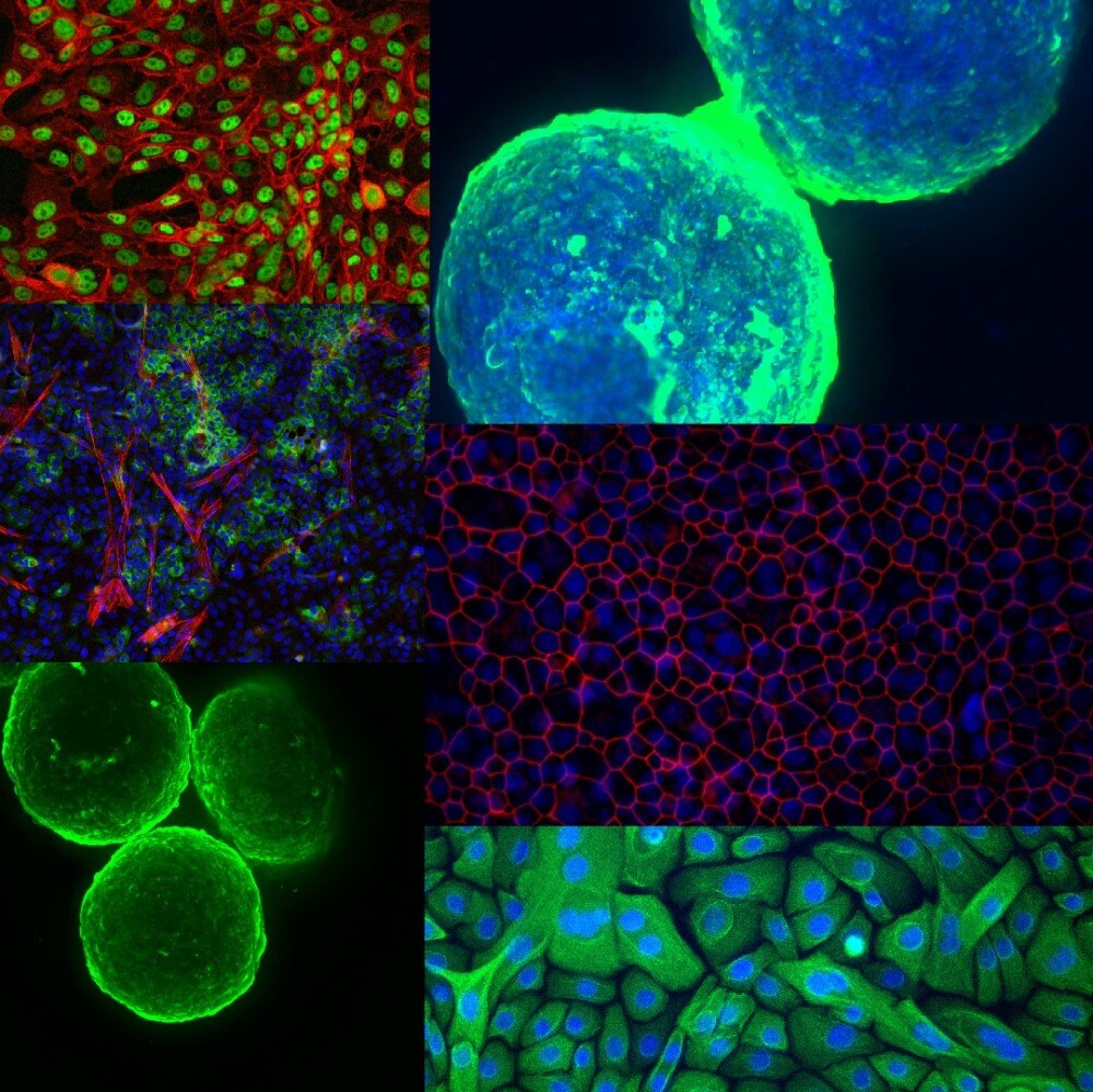
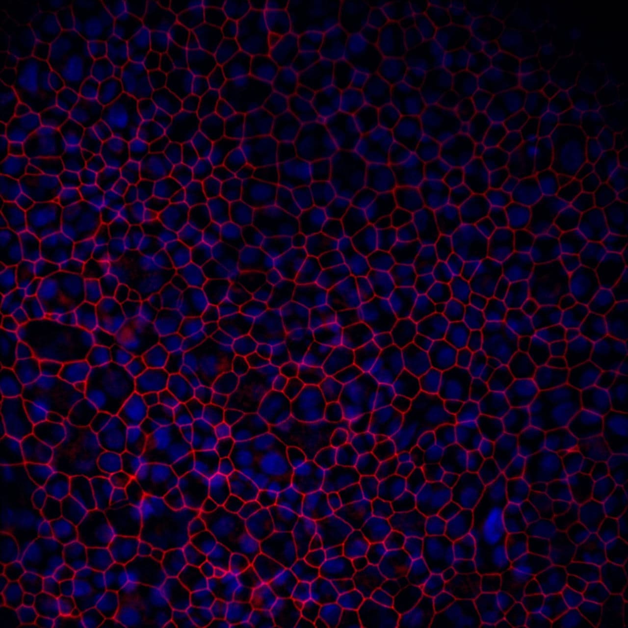
3. Which of the images from your research is most memorable to you?
The best memory is linked to this image where the tight junctions between bronchial epithelial cells can be seen. The reason is that this image was the first time that PMI could observe the composition of the 3D bronchial tissues we routinely use to assess our product. During my postdoc, I have indeed spent some time in testing various methods to stain the tissues and have ended up with a good protocol that allowed me to obtain this image and all the other ones.
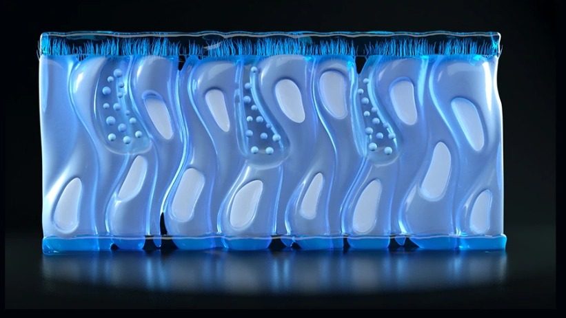
4. What was your favorite technique to use on the cells, cell cultures and spheroids shown on the images?
My favorite technique, which is also based on my main activity at PMI, is the development of 3D cultures that are further integrated into chip devices. Indeed, I find it mesmerizing to think that thanks to the current technologies available, we can partially recreate the complexity of the human body by combining 3D tissues created in the laboratories with an engineered microenvironment. This so called multi-organs-on-a-chip technology is fairly new and I am really happy that my job allows me to work on such devices.