The science we do at PMI covers a wide range of subjects, from airway deposition to genotoxicity analysis, and more. Within that scope, there are many moments of visual beauty that can capture the eye and increase our understanding of the field. Here, we've collected some images to offer more insight into the beauty of science, and how that beauty lets us learn more about an array of outcomes to help grow the global database of research.
Science is Beautiful
Science is Beautiful
Beakers, bunsen burners, and microscopes are the images that often come to mind while thinking about 'science', but there's obviously much more to the field than that when it comes to imagery.
Science is Beautiful
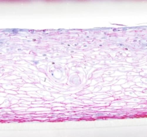
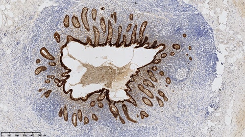
Pink Buccal
Magnified 20 times, this 3D human organotypic buccal #cell culture consists of normal, human-derived oral epithelial cells. Red-stained E-cadherin, a calcium-dependent adhesion molecule, acts as an adhesion receptor in the adherens junctions. Brown-stained Ki67 as a proliferation marker is an excellent marker to determine the growth fraction of a given cell population.
Science is Beautiful
Cytoskeletal Component
Vimentin, in brown in this human kidney tissue section, is the major cytoskeletal component of mesenchymal cells. As an intermediate filament protein, it is a member of the intermediate filament family.
Science is Beautiful
Spot the blue spots
At a closer look one can spot the blue spots. Those are cell nuclei in this 3D human organotypic buccal cell culture. The elegant brown E-cadherin is a calcium-dependent adhesion molecule and acts as an adhesion receptor in the adherens junctions, with important functions in cell adhesion and cell signaling.
Science is Beautiful
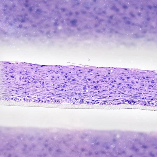
Vibrant Violet
Within this 3D human organotypic buccal cell culture, hematoxylin stains the nuclei blue while eosin stains the extracellular matrix and cytoplasm in varying degrees of pink. The vibrant violet cell culture consists of normal, human-derived oral epithelial cells and we are seeing the image magnified 20 times.
Science is Beautiful
Interview with Dr. David Bovard
"The most striking aspect of science and therefore of the nature that surrounds us is certainly the incredible work done by natural selection…"
David Bovard, Scientist – Microphysiological Systems, shares the aspects of science that are beautiful to him. He observes unexpected beauty in his research and has captured these beautiful images of cells, cell cultures and spheroids.
Science is Beautiful
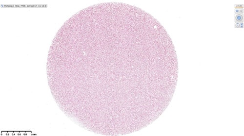
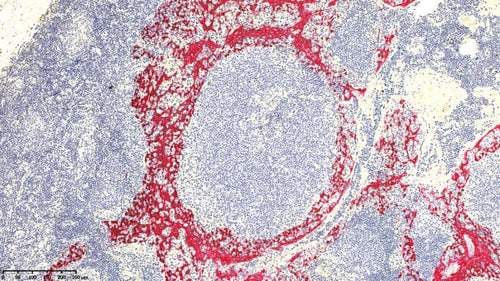
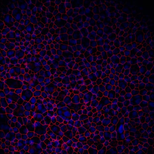
Circular Scope
This circular HeLa cell culture (comprised of immortal cells) acts as a positive control for RNAScope® technology – an in situ hybridization assay that detects RNA expression of target genes in various human cell cultures. Here, the target gene expression, human peptidylprolyl isomerase B, a "housekeeping gene" involved in basic cellular function is shown in red.
Science is Beautiful
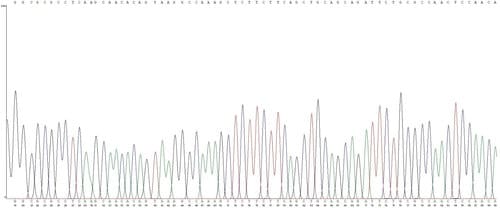
A Sequence of Plants
Sanger sequencing is used to show the A, T, C, G nucleotide sequence of DNA (developed by Nobel prize winner Frederick Sanger). Here, Sanger sequencing is used to show the tobacco plant DNA sequence as a chromatogram. Higher peaks correspond to higher intensity fluorescence signal.
Science is Beautiful
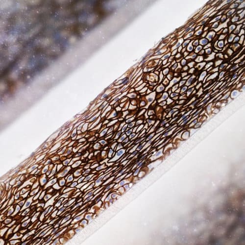
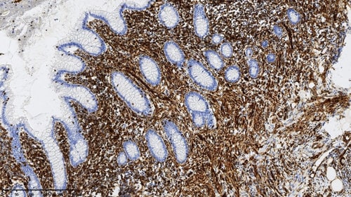
Adhesive Appendix
Immunohistochemical (IHC) staining of this human appendix tissue section marks E-cadherin molecules in brown. E-cadherin is a calcium-binding-dependent cell adhesion molecule – essentially it helps cells bind to other cells. Hematoxylin is used as a counterstain to contrast cell nuclei in blue.
Science is Beautiful
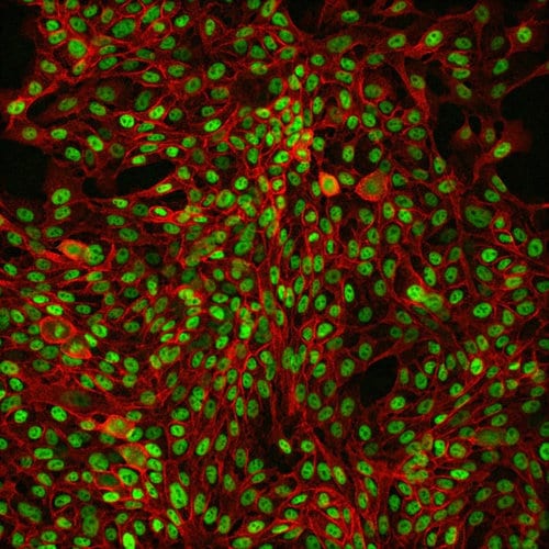
IHC Tonsil Tissue
This human tonsil tissue IHC staining colors the basal epithelial cell marker p63 and cytokeratin 5 (CK5) in brown, while cytokeratin 14 (CK14) is colored red. CK5 is seen as brown membranous staining, while p63 staining is found in the nuclei. Where p63 is absent in the nuclei a blue hematoxylin counterstain is seen.
Science is Beautiful
Intermediate
Regions of brown staining unveil the protein vimentin in this section of human appendix tissue. Vimentin is an intermediate filament protein that’s a major component of the cytoskeleton of mesenchymal cells.
Science is Beautiful
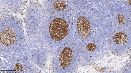
Ki67
Immunohistochemical staining in brown shows the protein Ki67, a marker for cell proliferation (multiplication) found in B-cells (a type of white blood cells) in this human tonsil tissue. We use this to visualize the growth fraction in this cell population.
Science is Beautiful
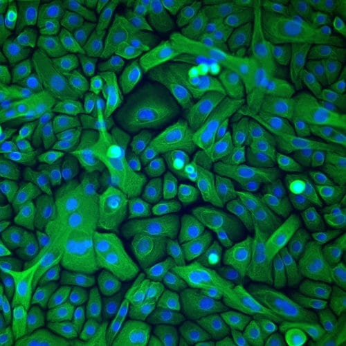
Cytoskeletal Green
These fluorescent cells have been stained green to show their cytokeratin 5, a protein that helps form the cytoskeleton. This primary human bronchial culture has been used to prepare 3D organotypic lung tissue for use in our organ-on-a-chip devices.
Science is Beautiful
Cytokeratin 5
Red, brown, and blue staining show CK5, p63, and cell nuclei, respectively. CK5 – also known as cytokeratin 5 – is a protein that helps to compose the filaments that create the cytoskeleton of the basal epithelial cells in this human tonsil tissue, p63 is a marker for these basal epithelia cells.
Science is Beautiful
Nasal Mucin
This 3D human organotypic nasal cell culture is stained to display Muc5AC (in brown) and nuclei (in blue). Mucus in the nasal pathway is made up of water, ions, and gel-forming mucin glycoproteins, among other components. Muc5AC is a mucin glycoprotein secreted by specialized goblet cells (which produce mucus).
Science is Beautiful
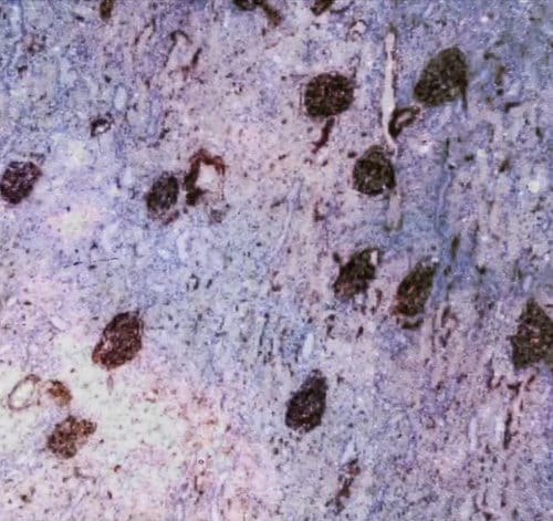
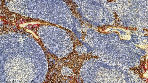
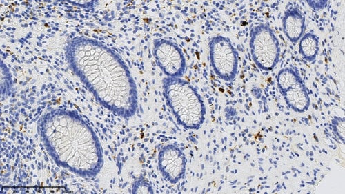
Gut IHC
CD68 is a protein expressed by macrophages and cells in the monocyte lineage (white blood cells). IHC staining in this appendix has colored the CD68 proteins in brown, while a hematoxylin counterstain identifies cell nuclei in blue.
Science is Beautiful
Fluorescent p63
This 3D nasal cell culture immunofluorescence staining displays the transcription factor p63 in green and nuclei in blue. The transcription factor acts as a sequence-specific DNA-binding transcriptional activator or repressor (it can stop or start the transcription of certain DNA sequences). In this 40x magnified image, p63 is used as a marker for basal cells.
Science is Beautiful
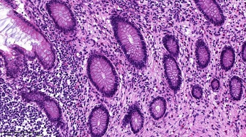
A Study in Indigo
Hematoxylin & eosin staining shows the mucosa (layers of cells containing mucous glands) of an appendix. From this indigo image you can see cross-sections of the glands – circular regions encapsulated in a darker purple.
Science is Beautiful
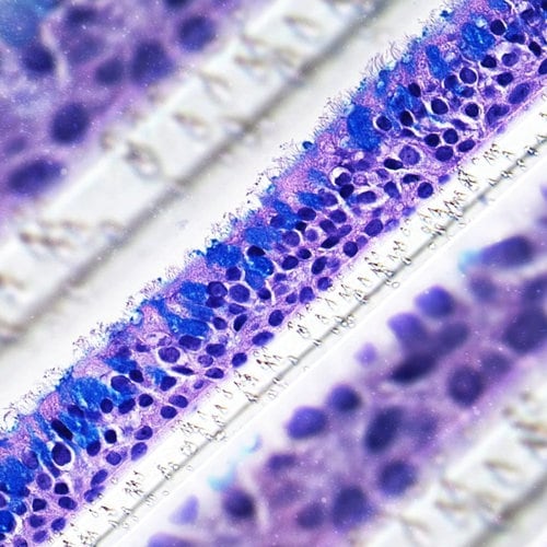
Culture of Violets
One of the most prominent tissue stains in histology (the study of microanatomy) is hematoxylin and eosin. Hematoxylin dyes the nuclei in this 3D human organotypic nasal cell culture blue, eosin dyes the extracellular matrix and cytoplasm pink, while the stain Alcian Blue dyes the mucin molecules (a component of mucus) blue.
Science is Beautiful
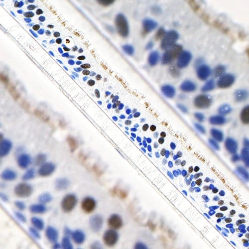
Nosy Fox
Within this 3D human organotypic nasal cell culture, immunohistochemical staining highlights the protein FOXJ1 in brown while hematoxylin stains the nuclei blue. The FOXJ1 protein is part of the forkhead box (Fox) family of transcription factors that play key roles in cell proliferation and differentiation.
Science is Beautiful
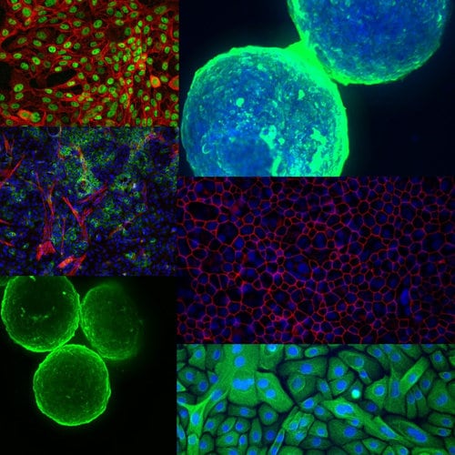
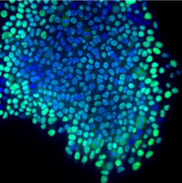
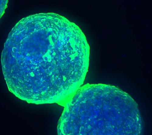
Undifferentiated
This speckled island of blue and green is a human induced pluripotent stem cell culture. These undifferentiated cells were cultured by one of our first R&D postdoctoral fellows for use in an in vitro neuronal model of Parkinson’s disease.
Science is Beautiful
Tricolore
Staining for albumin (green), nuclei (blue), and α-smooth muscle actin (red) of human liver cells and human hepatic stellate cells create this triple-colored façade. These 10x magnified cells were cultured together to develop a more complex model system of the human liver.
Science is Beautiful
Sticky Cells
This 3D human organotypic nasal cell culture shows blue-stained nuclei, and red-stained E-cadherin; a cell-cell adhesion molecule that plays a key role in cell behavior and tissue formation. 3D cell cultures more closely resemble the physiology of tissue in the human body, compared to 2D cultures or animal models.
Science is Beautiful
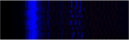
Electric Blue
How can we tell what DNA markers tobacco plants have? By separating the amplified microsatellite marker DNA with capillary electrophoresis, the process of loading DNA samples into gel capillaries and sending an electrical current through them. The current causes DNA molecules to travel through the gel, the smaller the DNA molecule the further away from the starting point they travel.
Science is Beautiful
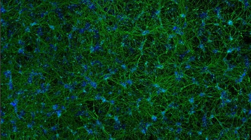
Emerald Neurons
The network of neuronal cells in this image have been derived from human induced pluripotent stem cells and stained in green, highlighting the neuronal marker tubulin beta III. Our research into neurons looks to establish a model of Parkinson’s disease, and test the potential neuroprotective effects of tobacco-derived compounds.
Science is Beautiful
Protein Junctions
In between these cells of a 3D human organotypic bronchial cell culture resides the protein ZO-1, stained in red. The protein is expressed in tight junctions; seals between cells that form a permeability barrier. The intact red trails mean that there’s no cell damage. In cases of cell/tissue damage, the tight junctions would not be visible.
Science is Beautiful
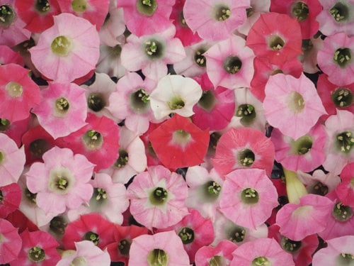
Pale to Pink
Believe it or not tobacco plants don't just produce leaves, they create a beautiful color palette of flowers. Our scientists are investigating flower diversity, starting with 'grandparents' that had pale or dark pink petals and cross-breeding those plants to produce offspring with the colors you see in the image.
Science is Beautiful
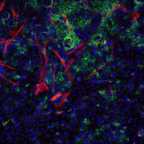
Bronchial Bloom
This picture highlights a slide of primary human bronchial cells - from the surface of the windpipe. The bright greens and reds in this image are created by staining for P63 and CD151 respectively. Why are we highlighting these cells? Because studying them gives us deeper knowledge of lung physiology - better understanding of their structure, function, and how they react to various stimuli.
Science is Beautiful
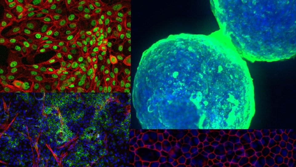
Luminous Spheres
These strange glowing orbs are human liver cell spheroids. But what are spheroids? They're a collection of cells used to mimic an organ. In our in vitro research we've developed lung/liver-on-a-chip technology for pre-clinical research. With further development, spheroids could potentially help supplement animal testing.
Science is Beautiful
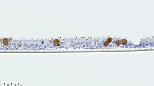
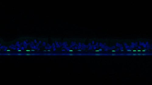
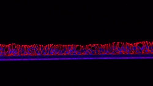
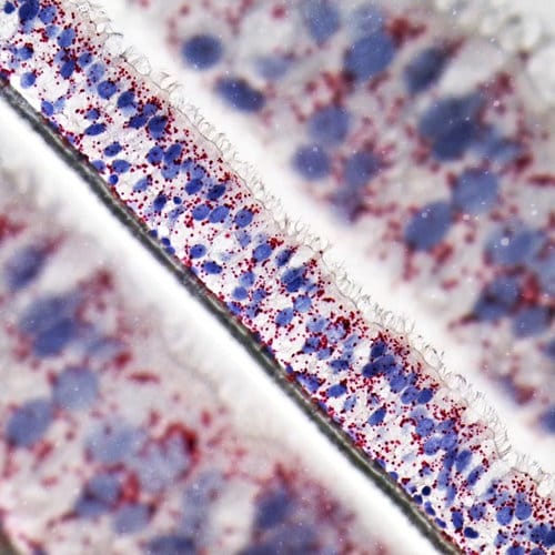
Nasal Housekeeping
This row of 3D human organotypic cells comes from nasal epithelium (inside a nose) made up of ciliated cells (which are topped with hair-like structures), goblet cells (which produce mucus), and basal cells (stem cells that can differentiate into other specialized cells). The flecks of red show the RNA expression of a gene called Hs-PPIB; a housekeeping gene.
Science is Beautiful is a project aimed at revealing some of the most beautiful aspects of the science we do at PMI. We’re proud of the 930+ scientists, engineers, technicians, and support staff working with us, state-of-the-art technologies we use, and the beauty of the science we do each day. If these images have piqued your interest, be sure to stay tuned on PMIScience.com and follow us on social media.